Brain Development Charts
Oct 13th 2022
Thanks to collaboration and Creative Commons Philosophy, I can share this great meta-project!
All authors recognition is to Reference: Bethlehem, R., Seidlitz, J., White, S. R. et al (2022). Brain charts for the human lifespan. Nature, 604(7906), 525–533.
https://doi.org/10.1038/s41586-022-04554-y
@ Long story short
An succesfull effort to conglomerate Brain Image Postprocessing Datasets, allows to plot Brain Charts for different Brain Develop Measurements along all the human lifespan and compare Controls Normals with Disease patients. "Currently, BrainChart is not a diagnostic tool, and therefore the resultant centile scores should only be used for research purposes."
Using body Grow Charts a Child is controlled by the Pediatrician. Healthy trajectories and also desviations (percentiles) from normal are detected.
Think in this concept, and by analogy apply it in Brain development. Ok, that is a Brain Chart for the entire Lifespan Development.
The data source are structured databases of fetal to old age MRI, postprocessed for specific targets like cortical Gray Matter Volume, White Matter Volume or subcortical Gray Matter Volume, and other measures.

FreeSurfer software is used to postprocess, and a standard template is uploaded and curated, from diverse Neuroimaging databases.
 Brainchart Site, its awesome!!
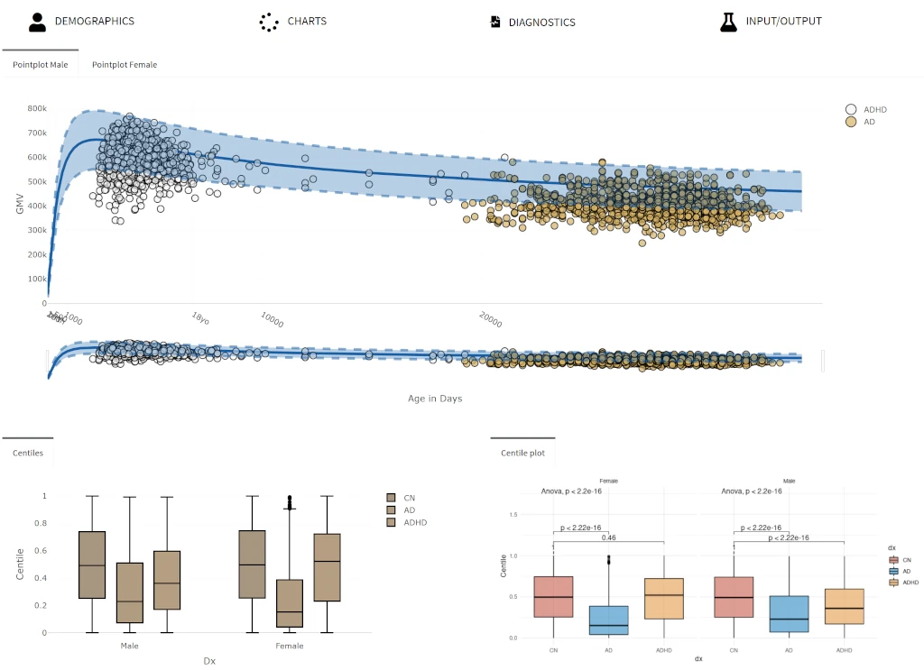
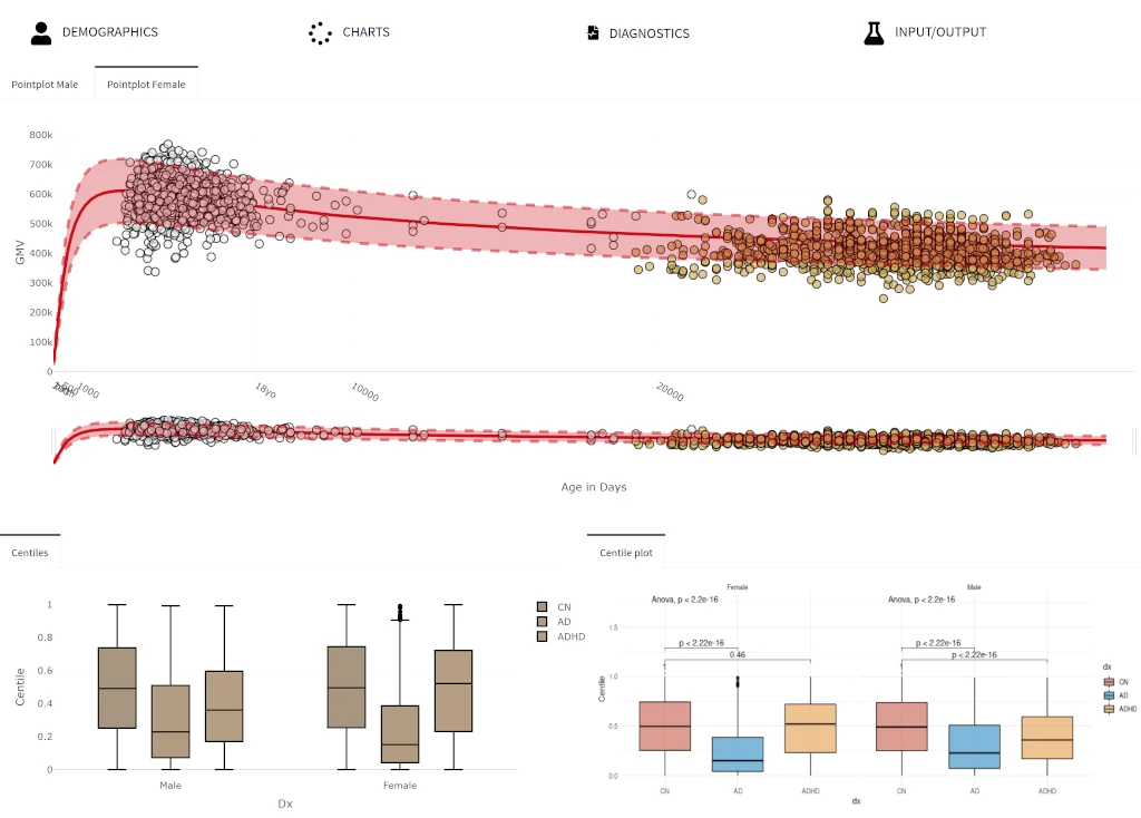
In this example, I use the online BrainChart to compare the Cortical Gray Matter Volume of the Brain in Lifespan, Normal controls (CN), Alzheimer Disease (AD) and Attention-Deficit/Hyperactivity Disorder (ADHD):
Click in each image to view in another window with a better resolution.
Caution!, remember that the advice is to not consider BrainCharts as a clinical tool. More research is needed.
(But, yes, in this big dataset of Brain MRI withFree-Surfer post processed analysis, the results shows a statistically significant lower Cortical Brain Gray Matter in AD and ADHD versus normal controls.)
All the code is written in R, this is the gitHub repository. Under a Creative Commons Attribution-NonCommercial-NoDerivatives 4.0 International License.
I promise to post about "when, how and why" I use: FreeSurfer, BrainStorm3, EDF Browser, Gephi, DSI-studio, SSPM, MicroDicom, Slicer, PSPP, Bioimage Suite Web and The Virtual Brain. And all the equivalent efforts in Python, if it exists.
For now, I refer you to this key resources:
The great NeuroImaging Tools & Resources Collaboratory site
The Neuroscience Information Framework.
The Center for Brains, Minds, and Machines
A very useful medical and health related sciences glossary: MeSH (Medical Subject Headings)


